Mast cells are cells of the immune system of the hematopoietic lineage that remain widely distributed in vascularized tissues throughout the body.
- Mast cells, unlike other immune cells, do not remain in the blood but remain localized in mucosal and epithelial tissues throughout the body.
- These are among the first cells to interact with environmental antigens and invading pathogens or microorganisms. Mast cells are thus considered one of the first cells of the immune system that interact with external antigens.
- Mast cells are primarily involved in the IgE-dependent responses, but these can also respond to a variety of other innate signals from pathogens, complement compounds, and other host cell types.
- Mast cells are also known to express many costimulatory molecules with immunoregulatory activities that help regulate the immune responses in the body.
- These are produced from the bone marrow and are released into the blood as undifferentiated cells. These cells then mature after they leave the blood and reach the specialized tissues.
- In the bone marrow, mast cells originate from CD34+/CD117+ pluripotent progenitor cells, and the maturation of these cells depends on KIT activation.
Structure of Mast Cells
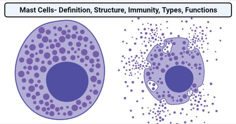
- Mast cells are mononuclear cells consisting of small secretory granules that range in size from 0.2 to 0.8 micrometers. In some cells, the granules are dense enough to obscure the appearance of the nucleus.
- The cells are oval or irregularly shaped with a single central nucleus. Within the nucleus, densely packed peripheral chromatin can be observed.
- Most of the cytoplasm is occupied by cytoplasmic granules but few secondary lysosomes. The cells also have small, finger-like projections from the cell membrane.
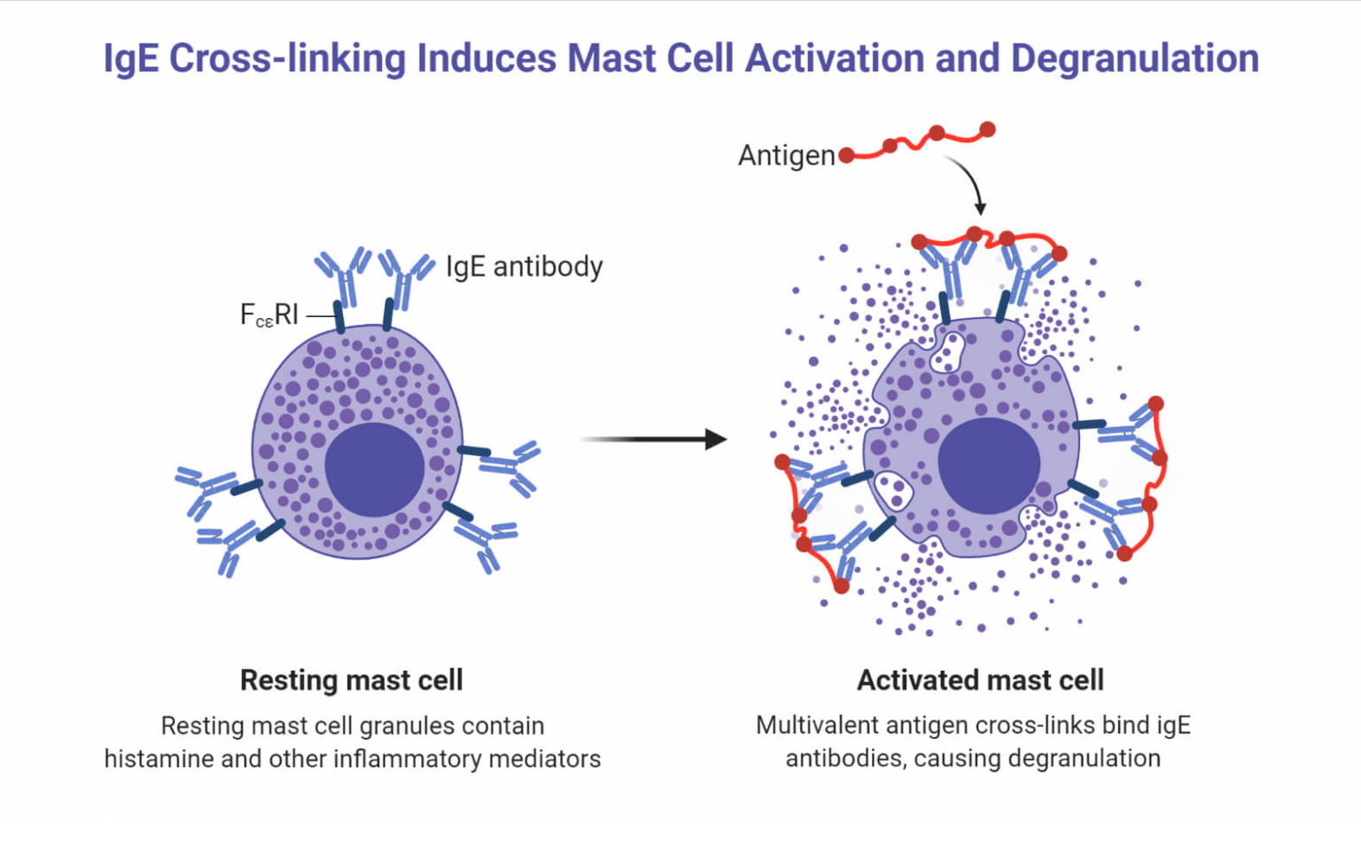
- The granules store inflammatory mediators like histamine, herapin, cytokines, and proteases.
- The plasma membrane of the mast cells has IgE receptors that bind to the Fc region of circulation IgE antibodies in order to induce degranulation of these mast cells.
- Mast cells share many features with basophils, but these can be distinguished based on their mononuclear morphology and monoclonal antibodies.
- Mast cells exist in three distinct forms based on their structure; intact cells, spreading cells, and degranulated cells.
- Intact cells are mostly found in blood vessels as well as in dermal tissues. The granules in these cells are closely packed together, which prevents the observation of other cellular organelles. The cells are usually spindle-shaped.
- Spreading cells are cells that occur in the superficial connective tissue. The granules are fewer in number than in the intact cell type.
- Degranulated cells are not metachromatic and have clear cytoplasm with a prominent nucleus.
Types of Mast Cells
- Mast cells in human beings can be differentiated into two types; mucosal and connective tissue mast cells.
- The connective tissue mast cells are found primarily in the loose connective tissue and skin, but these can be found in other connective tissues as well.
- The mucosal type mast cell is found in the gastrointestinal mucosa and peripheral airways.
- The exact factor that causes the differentiation of mast cells into the two types is not yet completely known.
- However, both of these types of mast cells can be activated by allergens and other nonallergic signals. Both types also have the IgE receptors responsible for the activation of degranulation of the cells.
- The difference between the two mast cells can be observed in their response to nonallergic signals, the mediators and their release, their proteoglycan constituents, and the makeup of the granular enzymes.
- The distinction between the two types of cells can be made easily in the case of rodents as these stains differently differ in size and are found in different tissues.
- The differentiation between the mast cells in humans can be made through the presence of chymase, a chymotryptic protease.
- The protease is only present in connective tissue mast cells, but the enzyme tryptic protease or tryptase is found in all human mast cells.
Functions of Mast Cells
The following are some of the functions of mast cells in the body;
- Mast cells secrete different pro-angiogenic factors like VEGF, bFGF, TGF-beta, and IL-8. The cells also release proteases and heparin, which induce permeability of the microvasculature, inducing angiogenesis.
- Mast cells are among the first line of defense against antigens entering the body. These cells are thus important for maintaining the homeostasis of the body, especially the gastrointestinal tract.
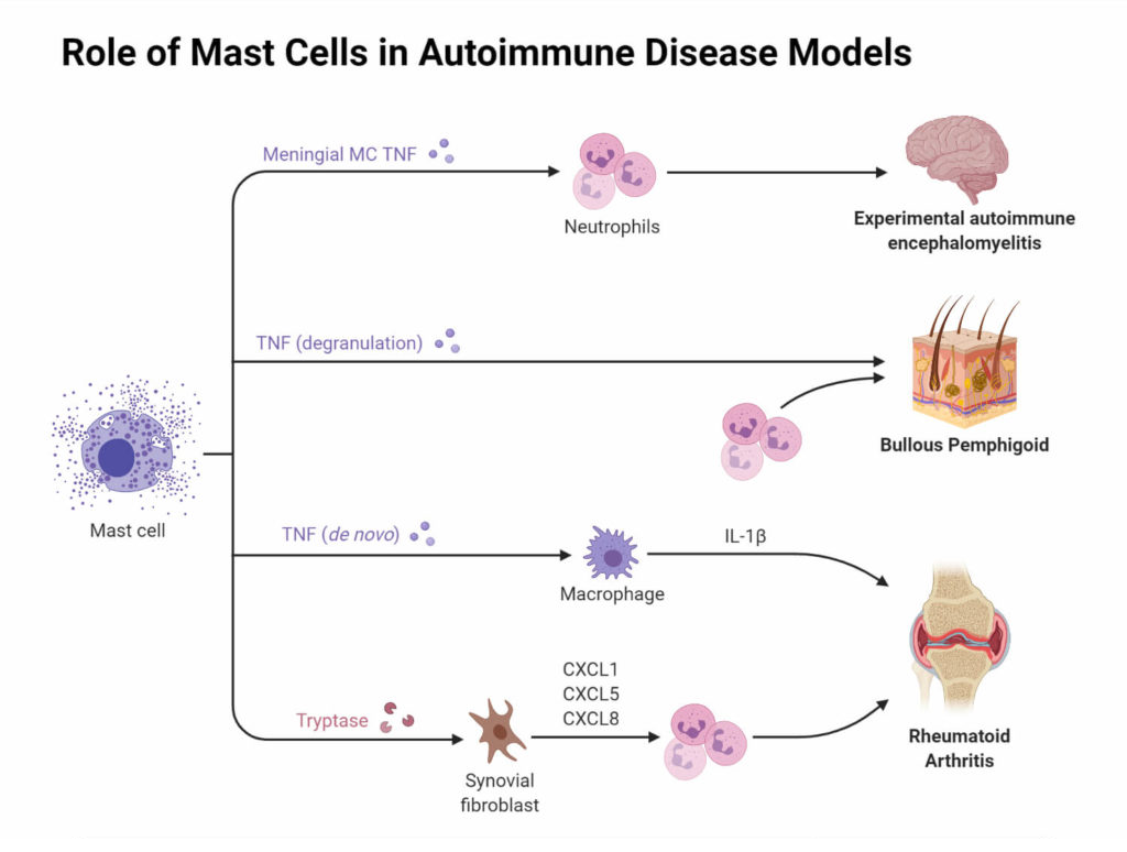
- In innate immunity, receptors on mast cells bind to antigens, causing a release of inflammatory mediators like IL-4, TNFα, and IL-6.
- These cells also help in providing immunity against viral antigens by the release of IFN-α and IFN-β.
- In adaptive immunity, mast cells process and present antigen via MHCI and MHCII, and these also activate dendritic cells, which are professional antigen-presenting cells.
- The most important and prominent function of mast cells is the release of newly synthesized mediators necessary for the immune response.
- Mast cells are also involved in an allergic reaction where the over-activation of mast cells can result in hyper-allergic reactions.