Microfilaments Definition
- Microfilaments, also called actin filaments, are polymers of the protein actin that are part of a cell’s cytoskeleton.
- They are long chains of G-actin formed into two parallel polymers twisted around each other into a helical orientation with a diameter between 6 and 8nm.
- Common to all eukaryotic cells, these filaments are primarily structural in function and are an important component of the cytoskeleton, along with microtubules and often the intermediate filaments. They are the smallest filaments of the cytoskeleton.
- Their functions include cytokinesis, amoeboid movement and cell motility in general, changes in cell shape, endocytosis and exocytosis, cell contractility and mechanical stability.
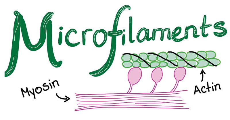
Figure: Diagram of Microfilaments
Structure of Microfilaments
- Microfilaments are primarily composed of polymers of actin, but in cells are modified by and interact with numerous other proteins.
- When actin is first produced by the cell, it appears in a globular form. But in microfilaments, however, they appear as long polymerized chains of the molecules are intertwined in a helix, creating a filamentous form of the protein i.e. F-actin.
- They are thus composed of two strands of subunits of the protein actin wound in a spiral. Specifically, the actin subunits that come together to form a microfilament are called globular actin (G-actin), and once they are joined together they are called filamentous actin (F-actin).
- They are usually about 7 nm in diameter making them the thinnest filaments of the cytoskeleton.
- The polymers of these linear filaments are flexible but still strong, resisting crushing and buckling while providing support to the cell.
- Like microtubules, microfilaments are polar. Their positively charged, or plus end, is barbed and their negatively charged minus end is pointed.
- Polarization occurs due to the molecular binding pattern of the molecules that make up the microfilament. Also like microtubules, the plus end grows faster than the minus end.
- Overall, they have a tough, flexible framework that helps the cell in movement.
- Microfilaments are typically nucleated at the plasma membrane. Therefore, the periphery (edges) of a cell generally contains the highest concentration of microfilaments.
- A number of external factors and a group of special proteins influence microfilament characteristics, however, and enable them to make rapid changes if needed, even if the filaments must be completely disassembled in one region of the cell and reassembled somewhere else.
- When finding directly beneath the plasma membrane, microfilaments are considered part of the cell cortex, which regulates the shape and movement of the cell’s surface.
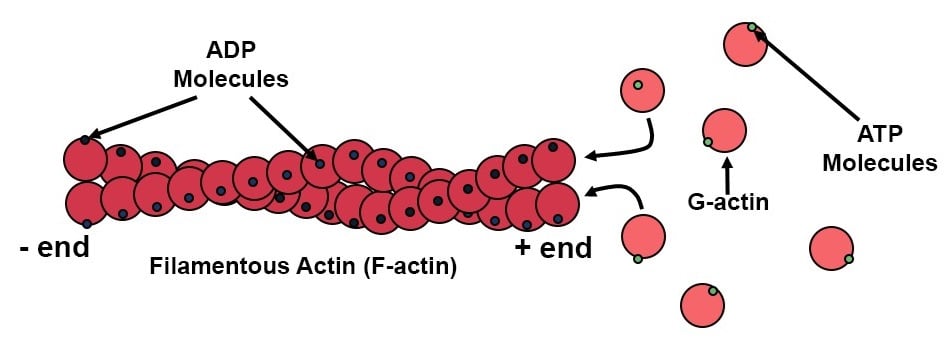
Formation or Self-assembly of Microfilaments
A microfilament begins to form when three G-actin proteins come together by themselves to form a trimer. Then, more actin binds to the barbed end. The process of self-assembly is aided by autoclampin proteins, which act as motors to help assemble the long strands that makeup microfilaments. Two long strands of actin arrange in a spiral in order to form a microfilament.
Functions
- In association with myosin, microfilaments help to generate the forces used in cellular contraction and basic cell movements.
- Eukaryotic cells heavily depend upon the integrity of their actin filaments in order to be able to survive the many stresses they are faced with within their environment.
- Microfilaments play a key role in the development of various cell surface projections including filopodia, lamellipodia, and stereocilia. The filaments are also hence involved in amoeboid movements of certain types of cells.
- Another important function of microfilaments is to help divide the cell during mitosis (cell division). Microfilaments aid the process of cytokinesis, which is when the cell “pinches off” and physically separates into two daughter cells.
- Microfilaments as a part of the cytoskeleton keep organelles in place within the cell. They provide cell rigidity and shape.
- They can depolymerize (disassemble) and reform quickly, thus enabling a cell to change its shape and move.

