Chromosomes structure are long, thread-like structures made of DNA and proteins that carry genetic information. They are located in the nucleus of eukaryotic cells and play a crucial role in heredity, cell division, and the regulation of genetic activity.
Structure of Chromosomes
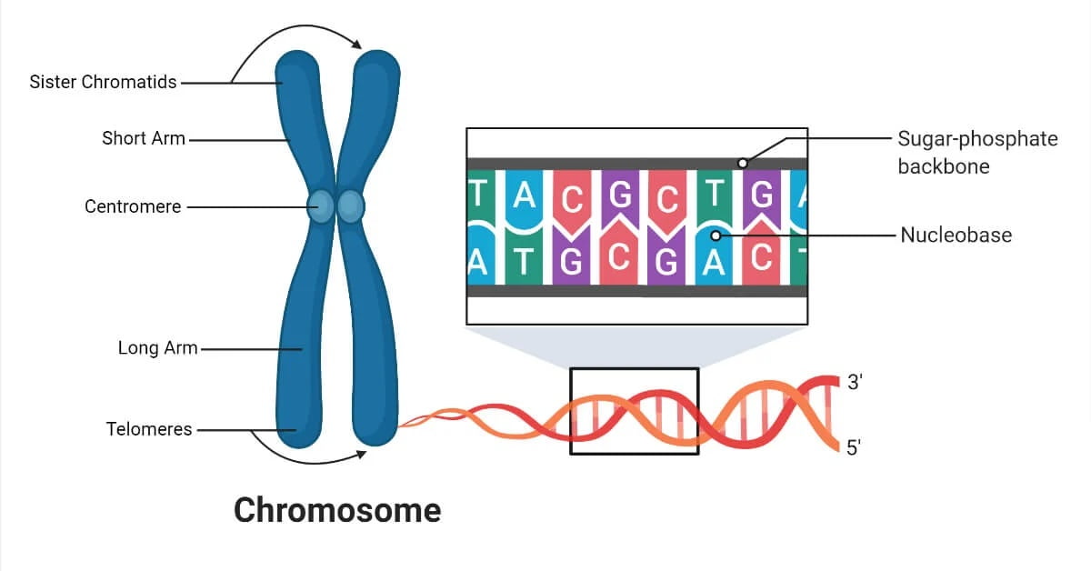
Basic Components: Chromosomes are composed of DNA tightly coiled around histone proteins, forming a structure known as chromatin. This arrangement helps in the efficient packaging of DNA and regulation of gene expression.
Histones and DNA Packing: Histones are basic proteins that assist in the organization of DNA into nucleosomes, the fundamental units of chromatin. Each nucleosome consists of DNA wrapped around a core of histone proteins, allowing the DNA to be compacted to fit inside the nucleus.
Chromosome Morphology:
Chromosomes have specific structural features, including:
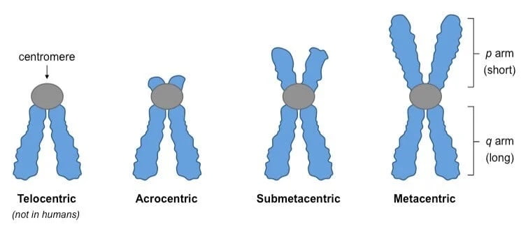
- Centromere: The constricted region of a chromosome, which plays a key role during cell division by holding sister chromatids together and attaching to spindle fibers. Centromeres can be positioned in different locations, leading to classifications such as metacentric (center), submetacentric (off-center), acrocentric (near one end), and telocentric (at the end).
- Telomere: The repetitive DNA sequences at the ends of chromosomes, protecting them from degradation and fusion with other chromosomes. Telomeres shorten with each cell division, playing a role in aging and cellular lifespan.
- Arms: The portions of a chromosome on either side of the centromere. Chromosomes have a short (p) arm and a long (q) arm. The length and structure of these arms vary between different types of chromosomes.
- Euchromatin: The less condensed form of chromatin that is transcriptionally active, meaning it contains genes that are being actively expressed. Euchromatin is found in the central parts of the chromosome arms and is important for gene regulation.
- Heterochromatin: The more condensed form of chromatin that is transcriptionally inactive, often containing repetitive sequences and structural components. Heterochromatin can be constitutive (permanently inactive) or facultative (inactive in certain cell types or developmental stages).
- Kinetochores: Protein complexes that form on the centromere and are crucial for chromosome movement during cell division. They attach to spindle microtubules and help in the accurate segregation of chromosomes.
- Satellite DNA: Repetitive, non-coding sequences found in specific regions of the chromosome, such as near centromeres (alpha-satellite DNA) and telomeres. Satellite DNA plays roles in chromosomal stability and function.
Types of Chromosomes Autosomes vs. Sex Chromosomes:
- Autosomes: Chromosomes that are not involved in determining the sex of an organism. Humans have 22 pairs of autosomes.
- Sex Chromosomes: Chromosomes that determine the sex of an organism. Humans have one pair of sex chromosomes (XX for females, XY for males).
Homologous Chromosomes: Homologous chromosomes are pairs of chromosomes (one from each parent) that have the same genes at the same loci but may carry different alleles.
Chromatid Structure: Each chromosome consists of two sister chromatids, which are identical copies formed during DNA replication and joined at the centromere.
Chromosomes and Cell Division
Chromosomes play a critical role in cell division, ensuring that genetic material is accurately replicated and distributed to daughter cells. This process is essential for growth, development, and maintenance of the organism.
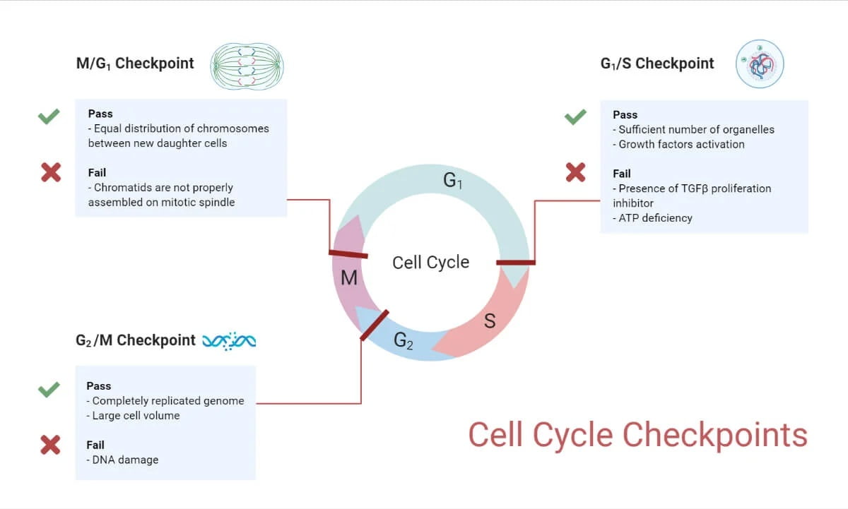
Overview of the Cell Cycle: The cell cycle is a series of phases that a cell goes through to grow and divide. It consists of interphase (G1, S, and G2 phases) and the mitotic phase (mitosis and cytokinesis).
Cell Division Cycle Phases
The cell cycle consists of interphase (G1, S, and G2 phases) and the mitotic phase (M phase). Here’s a detailed look at the content and amount of DNA and chromosomes during each phase:
G1 Phase (First Gap Phase):
- Content of DNA: The cell contains a normal diploid (2n) amount of DNA. For humans, this means 46 chromosomes, each consisting of a single chromatid.
- Amount of DNA: Each chromosome has one DNA molecule. In total, the cell has 2n DNA content (where n is the haploid number of chromosomes).
S Phase (Synthesis Phase):
- Content of DNA: DNA replication occurs during this phase, resulting in each chromosome consisting of two sister chromatids.
- Amount of DNA: The amount of DNA doubles from 2n to 4n. Although the number of chromosomes remains the same (46 in humans), each chromosome now has two DNA molecules (sister chromatids).
G2 Phase (Second Gap Phase):
- Content of DNA: The cell has completed DNA replication and now contains chromosomes with two sister chromatids.
- Amount of DNA: The cell maintains 4n DNA content. Each of the 46 chromosomes consists of two sister chromatids, so the total amount of DNA is twice the amount present in the G1 phase.
M Phase (Mitotic Phase): The M phase includes mitosis (prophase, metaphase, anaphase, telophase) and cytokinesis. Here’s a breakdown of DNA and chromosome content during these stages:
- Prophase:
- Content of DNA: Chromosomes condense and become visible, each consisting of two sister chromatids.
- Amount of DNA: The cell has 4n DNA content.
- Metaphase:
- Content of DNA: Chromosomes align at the metaphase plate, still consisting of two sister chromatids.
- Amount of DNA: The cell maintains 4n DNA content.
- Anaphase:
- Content of DNA: Sister chromatids separate and move to opposite poles of the cell.
- Amount of DNA: The separated chromatids are now considered individual chromosomes, so the cell temporarily has 4n chromosomes and 4n DNA content.
- Telophase:
- Content of DNA: Chromosomes reach the poles and begin to decondense. Nuclear envelopes reform around each set of chromosomes.
- Amount of DNA: The cell has 4n DNA content, which will be equally divided between the two daughter cells.
- Cytokinesis:
- Content of DNA: The cytoplasm divides, resulting in two separate daughter cells.
- Amount of DNA: Each daughter cell has 2n chromosomes and 2n DNA content, identical to the original parent cell before DNA replication.
In summary:
- G1 Phase: 2n DNA content, 2n chromosomes
- S Phase: DNA content increases from 2n to 4n, 2n chromosomes
- G2 Phase: 4n DNA content, 2n chromosomes
- M Phase:
- Prophase to Metaphase: 4n DNA content, 2n chromosomes
- Anaphase: 4n DNA content, 4n chromosomes (temporarily)
- Telophase and Cytokinesis: 2n DNA content, 2n chromosomes in each daughter cell
Mitosis Cell Division Cycle
Importance of Mitosis: Mitosis is a type of cell division that produces two genetically identical daughter cells from a single parent cell. It is essential for growth, tissue repair, and asexual reproduction in multicellular organisms.
Overview of Mitosis Stages: Mitosis is divided into five stages: prophase, metaphase, anaphase, telophase, and cytokinesis.
Mitosis Cell Cycle Steps
Prophase:
- Chromosomes condense and become visible.
- The nuclear envelope breaks down.
- Spindle fibers begin to form.
- Content of DNA: Chromosomes condense and become visible, each consisting of two sister chromatids.
- Amount of DNA: The cell contains 4n DNA content (double the DNA amount compared to the G1 phase).
Metaphase:
- Chromosomes align at the metaphase plate (center of the cell).
- Content of DNA: Chromosomes align at the metaphase plate, still consisting of two sister chromatids.
- Amount of DNA: The cell maintains 4n DNA content.
Anaphase:
- Sister chromatids are pulled apart to opposite poles of the cell by spindle fibers.
- Content of DNA: Sister chromatids are pulled apart to opposite poles of the cell, becoming individual chromosomes.
- Amount of DNA: The cell now has 4n chromosomes and 4n DNA content temporarily, as each chromatid is considered a separate chromosome.
Telophase:
- Chromatids reach the poles and decondense.
- Nuclear envelopes reform around the two sets of chromosomes.
- Content of DNA: Chromatids (now individual chromosomes) reach the poles and decondense. Nuclear envelopes reform around each set of chromosomes.
- Amount of DNA: The cell still has 4n DNA content, which will soon be divided between the two daughter cells.
Cytokinesis:
- The cytoplasm divides, resulting in two separate daughter cells.
- Content of DNA: The cytoplasm divides, resulting in two separate daughter cells.
- Amount of DNA: Each daughter cell has 2n chromosomes and 2n DNA content, identical to the original parent cell before DNA replication.
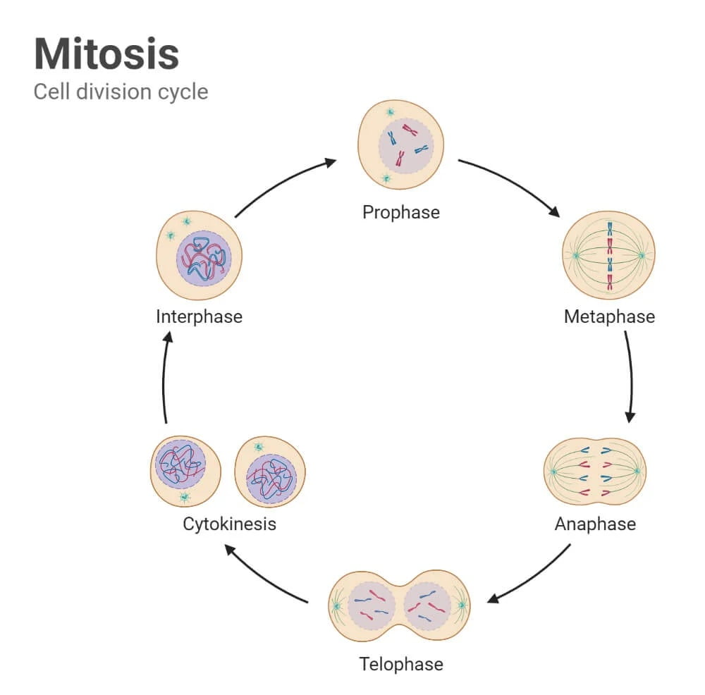
Summary of DNA Content and Chromosome Number During the Cell Cycle:
- G1 Phase: 2n DNA content, 2n chromosomes
- S Phase: DNA content increases from 2n to 4n, 2n chromosomes
- G2 Phase: 4n DNA content, 2n chromosomes
- M Phase (Mitosis):
- Prophase: 4n DNA content, 2n chromosomes
- Metaphase: 4n DNA content, 2n chromosomes
- Anaphase: 4n DNA content, 4n chromosomes (temporarily)
- Telophase and Cytokinesis: 2n DNA content, 2n chromosomes in each daughter cell
Chromosome Mutation
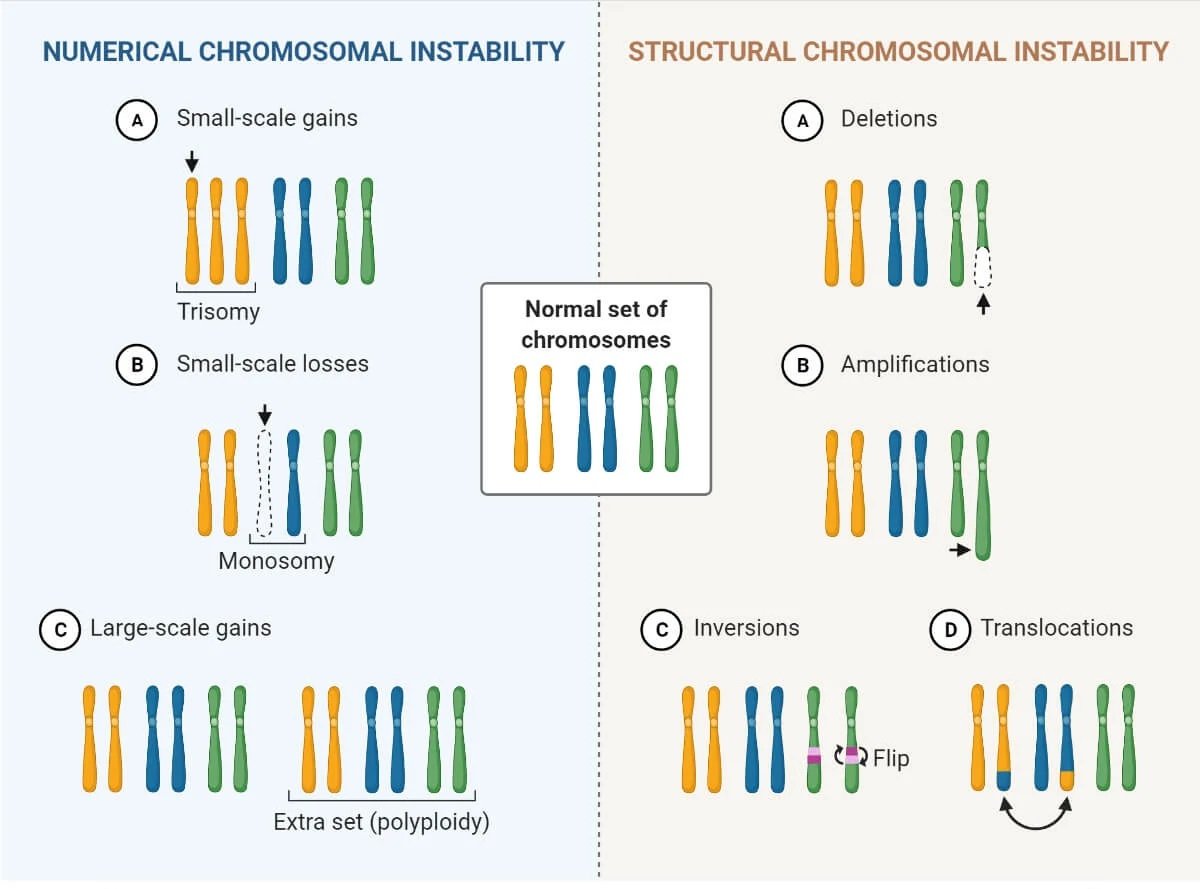
Definition and Significance: Chromosome mutations involve changes in the structure or number of chromosomes and can have significant effects on an organism’s phenotype.
Types of Mutations:
- Point Mutations: Changes in a single nucleotide.
- Deletions: Loss of a chromosome segment.
- Duplications: Repetition of a chromosome segment.
- Inversions: Reversal of a chromosome segment.
- Translocations: Rearrangement of parts between nonhomologous chromosomes.
Causes of Mutations in Chromosomes
Environmental Factors:
- Radiation: Exposure to ionizing radiation can cause DNA breaks and chromosomal aberrations.
- Chemicals: Certain chemicals (mutagens) can alter DNA structure and cause mutations.
Biological Factors:
- Errors in DNA Replication: Mistakes during DNA replication can lead to mutations.
- Viruses: Some viruses can integrate their genetic material into the host genome, causing mutations.
Types of Mutations
Mutation I: Gene Mutations: Changes in the nucleotide sequence of a single gene, affecting the function of the encoded protein.
Mutation II: Chromosomal Mutations: Changes in the structure or number of chromosomes, affecting many genes. Examples include deletions, duplications, inversions, and translocations.
Common Chromosome Disorders
Down Syndrome (Trisomy 21): Caused by an extra copy of chromosome 21, leading to developmental delays and physical characteristics such as a flat facial profile and almond-shaped eyes.
Klinefelter Syndrome (XXY): Affects males who have an extra X chromosome, leading to symptoms such as reduced testosterone levels, breast development, and infertility.
Turner Syndrome (Monosomy X): Affects females who have only one X chromosome, leading to short stature, webbed neck, and infertility.
Identification of Genetic Disorders
Karyotyping: A laboratory technique that visualizes chromosomes under a microscope, allowing the detection of numerical and structural abnormalities.
Molecular Techniques:
- PCR (Polymerase Chain Reaction): Amplifies specific DNA sequences for analysis.
- FISH (Fluorescence In Situ Hybridization): Uses fluorescent probes to detect specific DNA sequences on chromosomes.
Diagnosis of Chromosomal/Genetic Disorders
Prenatal Diagnosis:
- Amniocentesis: Sampling of amniotic fluid to analyze fetal chromosomes.
- Chorionic Villus Sampling (CVS): Sampling of placental tissue to analyze fetal chromosomes.
Postnatal Diagnosis:
- Newborn Screening: Tests performed on newborns to detect genetic disorders early.
- Genetic Testing: Analyzes an individual’s DNA to identify genetic mutations and diagnose genetic disorders.
Chromosome Functions
Gene Expression and Regulation: Chromosomes house genes that are transcribed into RNA and translated into proteins, controlling cellular functions and development.
Inheritance of Traits: Chromosomes carry genetic information from parents to offspring, determining inherited traits.
Maintaining Genetic Stability: Chromosomes ensure the accurate replication and segregation of genetic material during cell division, preserving genetic integrity.

