The cellular membranes of living organisms possess semi-permeability, allowing only specific ions, molecules, and water to pass through. The permeability of these molecules varies depending on the structural arrangement of the lipid bilayer found in different cell types and organisms.
The lipid bilayer, together with macromolecules like proteins, carbohydrates, and lipids, facilitates the transportation of molecules across the membrane. The membrane’s ability to regulate molecular transport is crucial for the organism’s life processes.
Two modes of transport are utilized by membranes for molecular movement:
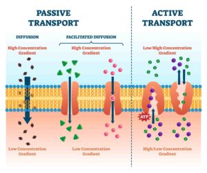
Passive Transport: This process involves the movement of substances across the membrane along the concentration gradient, from areas of high to low concentration, without the expenditure of energy.
Examples include the diffusion of neurotransmitters such as acetylcholine across neuronal synapses, the diffusion of gases in the lungs through alveolar membranes, and the movement of water molecules through osmosis.
Active Transport: In contrast, active transport involves the movement of molecules across the membrane against the concentration gradient, from areas of low to high concentration, requiring energy expenditure typically derived from ATP (adenosine triphosphate).
Examples include the expulsion of Ca2+ ions from cardiac muscle cells, the transportation of glucose molecules across membranes, and the movement of amino acids across the intestinal lining in the human gut.
- Semi-Permeable Cellular Membrane
- Allows only a fraction of ions, molecules, and water across
- Permeability varies based on lipid bilayer structure among different cells and organisms
- Lipid Bilayer
- Works with macromolecules (proteins, carbohydrates, lipids) to facilitate transport
- Essential for controlling transportation of molecules across the membrane
- Modes of Transport
- Passive Transport
- Movement of substances from high to low concentration
- No energy expenditure
- Examples:
- Diffusion of neurotransmitters (e.g., acetylcholine) across synaptic junctions in neurons
- Diffusion of gases in the lungs across alveolar membranes
- Movement of water molecules by osmosis
- Active Transport
- Movement of substances from low to high concentration
- Requires energy expenditure from ATP (adenosine triphosphate)
- Examples:
- Movement of Ca2+ ions out of cardiac muscle cells
- Transportation of glucose molecules across membranes
- Movement of amino acids across the intestinal lining in the human gut
- Passive Transport
Mutational alterations in the transportation system or the membrane transporters implicated can potentially result in significant health disorders, including depression, cystic fibrosis, drug abuse, migraine, and epilepsy.
Lipid Bilayers and Membrane Proteins
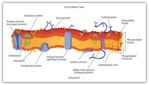
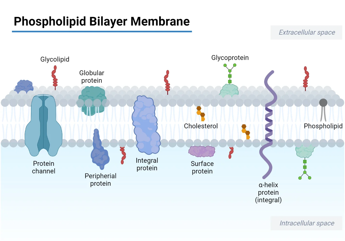
- Biological membranes typically consist of a phospholipid bilayer, where the hydrophilic side faces outward and the hydrophobic side inward, along with transmembrane proteins spanning the bilayer.
- These integrated proteins serve as channels, pumps, and carriers essential for transporting ions and macromolecules across biological membranes.
- Channel proteins create pores in the membrane, allowing the unimpeded passage of inorganic ions such as Na+, Cl-, K+, and H+, as well as molecules of appropriate size. These channel proteins are tightly regulated by extracellular signals, which govern the movement of ions and solutes across the membrane.
- Carrier proteins function akin to enzymes, selectively binding to and transporting specific small molecules like glucose to facilitate their translocation across the membrane.
- The ratio of phospholipids to proteins varies depending on the cell type. For instance, the mitochondrial inner membrane contains approximately 75% protein, indicating the presence of protein complexes involved in oxidative phosphorylation and the electron transport system.
Active Transport
Active transport refers to the energy-driven movement of ions, small molecules, and solutes across biological membranes against an electrochemical or concentration gradient, typically from lower to higher concentration.
This process requires cellular energy expenditure and plays a crucial role in maintaining the homeostasis of molecules and ions within cells.
Active transport can be categorized into two main types:
- Primary Active Transport: This type of active transport utilizes ATP (adenosine triphosphate) as an energy source to transport solutes against their concentration gradient.
- Secondary Active Transport: In secondary active transport, energy is derived from an electrochemical gradient established by primary active transport or other cellular processes. This energy is used to transport molecules against their concentration gradient.
Primary Active Transport
Primary Active Transport, also known as Direct Active Transport, involves the direct utilization of metabolic energy to transport substances across the membrane. This process relies on the breakdown of ATP (adenosine triphosphate) to ADP (adenosine diphosphate), releasing energy in the process.
This mode of transport is utilized for the movement of various metal ions such as K+, Na+, Cl-, H+, Mg2+, and Ca2+. The enzymes responsible for facilitating primary active transport of ions are ATPases, which actively transport charged ions across ion channels or pumps in the membrane.

Types of Primary Active Transport
P-Type ATPases
- Also known as E1-E2 ATPases due to their ability to switch between two conformations.
- Play diverse roles in humans, including nerve impulses, nutrient absorption in the intestine and kidney, and muscle relaxation.
- Found in eukaryotes and bacteria, these ion channels autophosphorylate using ATP and aspartate.
- Enzyme structure typically includes four functional domains:
- P-domain (Phosphorylation): Contains amino acids like aspartate.
- N-domain: Responsible for nucleotide binding.
- A-domain: Actuator domain with a phosphatase domain for dephosphorylation.
- R-domain: Regulatory domain.
- Examples include:
- Na+-K+ (sodium-potassium) pump
- Calcium ATPase (Ca2+ pump)
- Hydrogen ATPase (H+ pump)
- H+/K+ ATPase in proton-potassium pump in various organisms.
Mechanism of Sodium-Potassium (Na+/K+ ATPase) Pump:

- Uses ATP to change protein conformation, moving three Na+ ions out of the cell and two K+ ions into the cell.
- Ouabain blocks K+ influx by inhibiting protein dephosphorylation.
F-ATPase
- Found in mitochondrial inner membranes and chloroplast thylakoid membranes.
- Also known as ATP phosphohydrolase or ATP synthase.
- Uses rotating machinery powered by ATP hydrolysis to pump Na+ or H+ ions.
- Composed of two main domains:
- F1 Domain: Hydrophilic catalytic globular domain with an asymmetric hexameric ring and a central stalk, containing catalytic sites for ATP hydrolysis and synthesis.
- F0 Domain: Hydrophobic, embedded in the membrane, responsible for ion translocation.
- Drives ATP synthesis using energy from proton movement and functions as ATP synthase under low driving force conditions, generating a transmembrane electrochemical gradient.
V-ATPase
- Known as vacuolar ATPases, proton-translocating pumps in the tonoplast of cells, making organelles more acidic than the surrounding cytoplasm.
- Utilizes energy from ATP hydrolysis to transport protons across membranes.
- Multimeric protein with two domains:
- V1 Domain: Catalytic site for ATP hydrolysis, comprises eight subunits.
- V0 Domain: Responsible for proton movement across the membrane.
- Acts as a molecular switch between anabolic and catabolic metabolism, with structural disruptions leading to diseases like cancer.
ATP Binding Cassette (ABC) Transporters
- Found in chloroplasts, mitochondria, and plasma membranes.
- Involved in detoxification, phytohormone transport, and pathogen response.
- Utilize energy from ATP hydrolysis to transport solutes across membranes.
- Composed of four domains:
- Two transmembrane integral domains spanning the membrane six times.
- Two domains responsible for ATP hydrolysis.
- Export antimicrobial metabolites and volatile compounds such as benzene and 1,3-butadiene.
- In humans, approximately 48 genes associated with various diseases, including Dubin-Johnson Syndrome, Cystic Fibrosis, Ataxia, Anemia, and adrenoleukodystrophy.
- Participate in drug/antibiotic resistance, signal transduction, antigen presentation, and bacterial pathogenesis.
Secondary Active Transport
Also Known As: Coupled Transport or Cotransport.
- Mechanism: Involves the movement of ions along with larger molecules like glucose.
- Step 1: Ions are pumped down the electrochemical gradient (but against a concentration gradient), releasing energy.
- Step 2: Solute is translocated across a membrane down the concentration gradient.
Functions:
- Generates an electrochemical gradient due to the movement of ions at the expense of energy across a membrane.
- Ion gradient difference leads to solute movement in either:
- Antiport (opposite direction)
- Symport (same direction)
Ion Usage:
- Yeast and Bacteria: Commonly use hydrogen (H+) ions.
- Humans: Widely use sodium (Na+) ions.

Types of Secondary Active Transport
Based on Direction of Solute and Ions Across a Membrane:
- Antiport (Countertransport):
- Solutes and ions move in opposite directions.
- Examples:
- Na+/Ca2+ Antiporter: Regulates low calcium concentration in cardiac muscle cells, exchanging three Na+ ions down the gradient for one Ca2+ ion against the gradient (3:1 ratio).
- Cl-/HCO3- Antiporter: Provides electroneutral exchange, moving one Cl- ion down the gradient per bicarbonate (HCO3-) ion against the gradient (1:1 ratio).
- Na+/H+ Antiporter: Maintains cytoplasmic pH, transporting one Na+ ion down the gradient per H+ ion against the gradient (1:1 ratio).
- Symport (Cotransport):
- Solutes and ions move in the same direction.
- Examples:
- Glucose Symporter (SGLT1): Found in intestinal epithelium, kidney nephrons, brain, and heart. Imports two Na+ ions down the gradient per glucose molecule against the gradient.
- GABA Symporter (GAT): Regulates the concentration of GABA in the synaptic cleft, coupled with Na+ or Cl- ions.
- Oligopeptide Symporter (PepT): Crucial for drug entry (e.g., beta-lactam antibiotics) and nitrogen reabsorption in the intestine and kidney. Transports one H+ ion from high to low concentration per dipeptide or tripeptide molecule against the gradient.
Concentration Capacity:
- Measured by the ion/substrate coupling ratio.
- Example: Sodium (Na+) ions to glucose have a coupling ratio of 2:1, indicating two Na+ ions move for each glucose molecule per transport cycle.


